Skin Analysis Software Data Privacy: Clinic Guide
Skin analysis software data privacy starts with knowing where images go, why they are used, who can access them, and how each copy is removed....

A skin analyzer does not photograph a face and grade it by eye. It sends light into the skin, reads what comes back, and compares that signal with a reference baseline. Skin tone changes the signal long before any software draws a conclusion.
Most people expect a camera that scores a face. In practice, the chain has three steps. A light source illuminates the skin; a sensor records the light that returns; software turns that pattern into numbers and grades. Each step can shift when skin tone changes, which is why two clients with a similar-looking concern can leave with quite different reports.
When light reaches the skin, it follows three rough paths. Some reflects straight off the surface, and the amount depends on texture, oil and how the face is lit. Some travels into the epidermis and dermis, where melanin, hemoglobin and water absorb parts of it. The rest scatters and finds its way back to the camera.
Melanin absorbs across roughly 400–700 nm, with absorption falling as the wavelength grows. Hemoglobin absorbs strongly in the green-to-yellow part of the spectrum. Because those two pigments take different slices of the light, a camera can in principle tell them apart — but only if it captures them through different channels. When more melanin sits in the epidermis, less light reaches the deeper layers and less returns. A clinic can picture this as taking a photograph in a dim room: turning up the brightness helps, yet it also brings in noise.
The sensor records brightness and color, not “a wrinkle” or “a spot”. Software then compares those values with a reference range and decides where a feature sits. That reference range is the baseline, and the cut-off between unremarkable and notable is a threshold. Both are engineering choices made before the client sits down.
Clinicians often group skin tone with the Fitzpatrick phototypes, a public classification that sorts skin by how it responds to sun exposure. It is a shared vocabulary, not a measurement made by the device. Once a baseline has been chosen, the same pixel pattern can fall inside the range on one setting and outside it on another. For that reason, a score describes a comparison rather than a fact about the skin.
These outputs support consultation and progress tracking. They are not a diagnosis, and a practitioner should refer anything suspicious to a physician.
When a clinic compares one skin analysis machine with another, six parameters decide how fairly it reads across skin tones. None of them works in isolation.
| Parameter | What it describes | Why skin tone matters |
| Imaging channels | Which wavelengths and polarization states the device captures | Separate pigment and blood channels stop melanin from being counted as redness |
| Sensor and resolution | How much surface detail a single frame holds | Low-contrast texture needs detail when the return signal is already weak |
| Capture angles and positioning | Whether the face stays at a fixed distance and angle | Repeat scans compare like with like only when geometry stays put |
| Exposure and skin-tone baseline | How the camera sets brightness and reference levels before a shot | One fixed exposure can leave deeper skin under-lit and lighter skin washed out |
| Reading thresholds | Where the software draws the line between unremarkable and notable | A baseline set on lighter skin may flag ordinary pigmentation on deeper skin |
| Report outputs | Which values, grades and comparisons the report shows | Area, percentage and historical views let staff question a score instead of accepting it |
A higher number in any single row does not solve the problem. If a team raises resolution but keeps one fixed exposure, fine pigment detail on deeper skin still disappears into noise. Equally, a well-exposed image tells little when the software folds redness and pigment into one map. What matters is how the rows work together.
Once the light path and the scoring rules are clear, the effect of skin tone becomes easier to describe. It shows up in three places, and they often overlap.
More melanin means a weaker return signal. Under the same exposure, small differences in brightness — the edge of a pore, a faint patch of early pigment, a shallow line — shrink toward the noise floor. Staff may then see a “clean” image simply because the contrast was never captured. Overcorrecting brings the opposite fault: raising gain or illumination to recover detail can clip the highlights on lighter skin, so a different set of features vanishes. Either way the missing information is physical, not a software bug.
Pigment channels compare what they see with a reference. On deeper skin the ordinary melanin signal already sits high, even where there is no concern at all. If the reference was built around lighter skin, that normal signal crosses the threshold and the report calls it excessive pigmentation. The same thing happens when exposure or reference settings change between visits. The settings alter what the image looks like, while the face stays exactly as it was. Clients who notice this lose confidence fast, so it helps to explain the setup before showing the grade.
Melanin and hemoglobin absorb different wavelengths, yet a single broad-spectrum image renders both as a darker area. Without separate channels, a vascular flush and a pigment patch can be read as the same finding. Cross-polarized light suppresses surface glare and lets deeper color show through, whereas parallel-polarized light keeps surface texture visible. Under UV light, porphyrins give a brick-red fluorescence while melanin stays dark. Those contrasts are only available to a device that captures them separately.
Three common misreadings follow from this:
Teams that work with skin analysis equipment across a mixed client base usually write a short room checklist, because a steady routine achieves more than a clever correction applied afterwards.
Before the client sits down, staff can clean the skin and ask that makeup be removed. They should avoid scanning on the same day as an exfoliating treatment or an energy-based procedure, since both temporarily change surface reflection. Indirect lighting helps more than bright overhead spots, and the chair should sit away from a window with direct sun behind it.
During the shot, a forehead rest, a chin rest and a centered guide line hold the face at the same distance and angle. A hood or shroud keeps stray light out of the frame. The pigment channel and the blood channel are then read side by side. Cross-polarized views take care of oil glare.
For a follow-up visit, the same settings, the same time of day and the same capture angles matter as much as the treatment itself. Unless those stay fixed, a lower score may only record a brighter afternoon.
The practical value shows up most clearly in clinics whose clients span several skin tones. An aesthetics practice can build a baseline before a course of treatment, talk through the plan with images, and review the same area afterwards. Chain salons use before-and-after views to explain a facial treatment and to keep a member’s history in one place. Cosmetics counters and brand training teams compare sunscreen coverage before and after application, which turns an invisible claim into something a customer can see.
Communication is the part that often fails. When a report says only “pigmentation is high”, a client with deeper skin may hear a sales pitch rather than an observation. The reaction is stronger still when the flagged area is simply the natural color of a cheekbone. Once staff can point to one channel for melanin and another for blood, the conversation moves away from opinion. They can say which area is pigment, which is a flush, and which has changed since the last visit. That distinction is what makes a repeat scan worth doing at all.
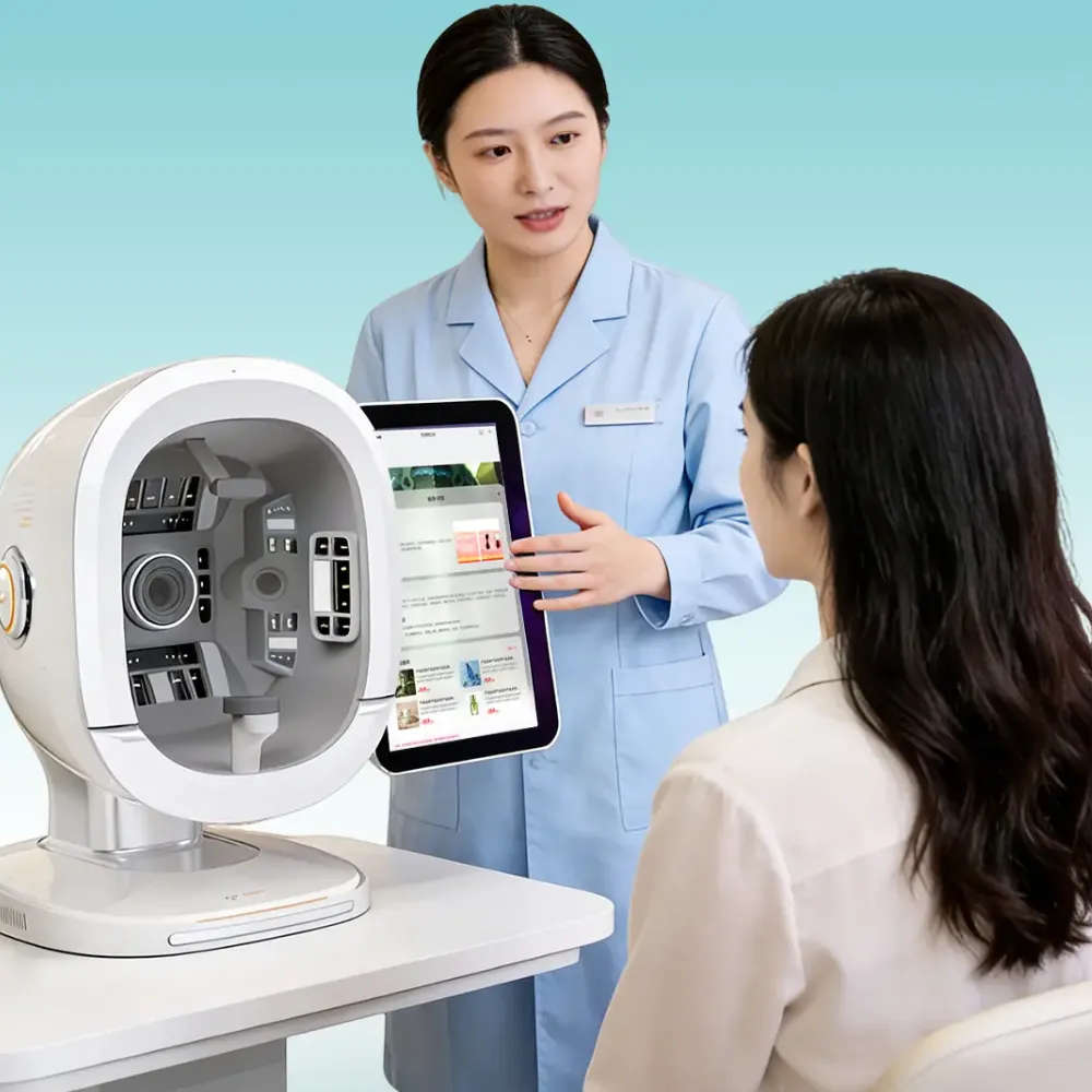
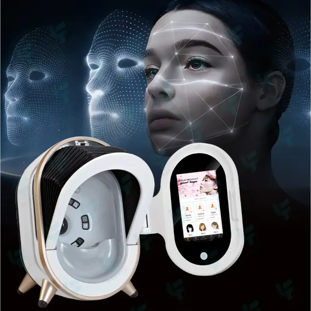
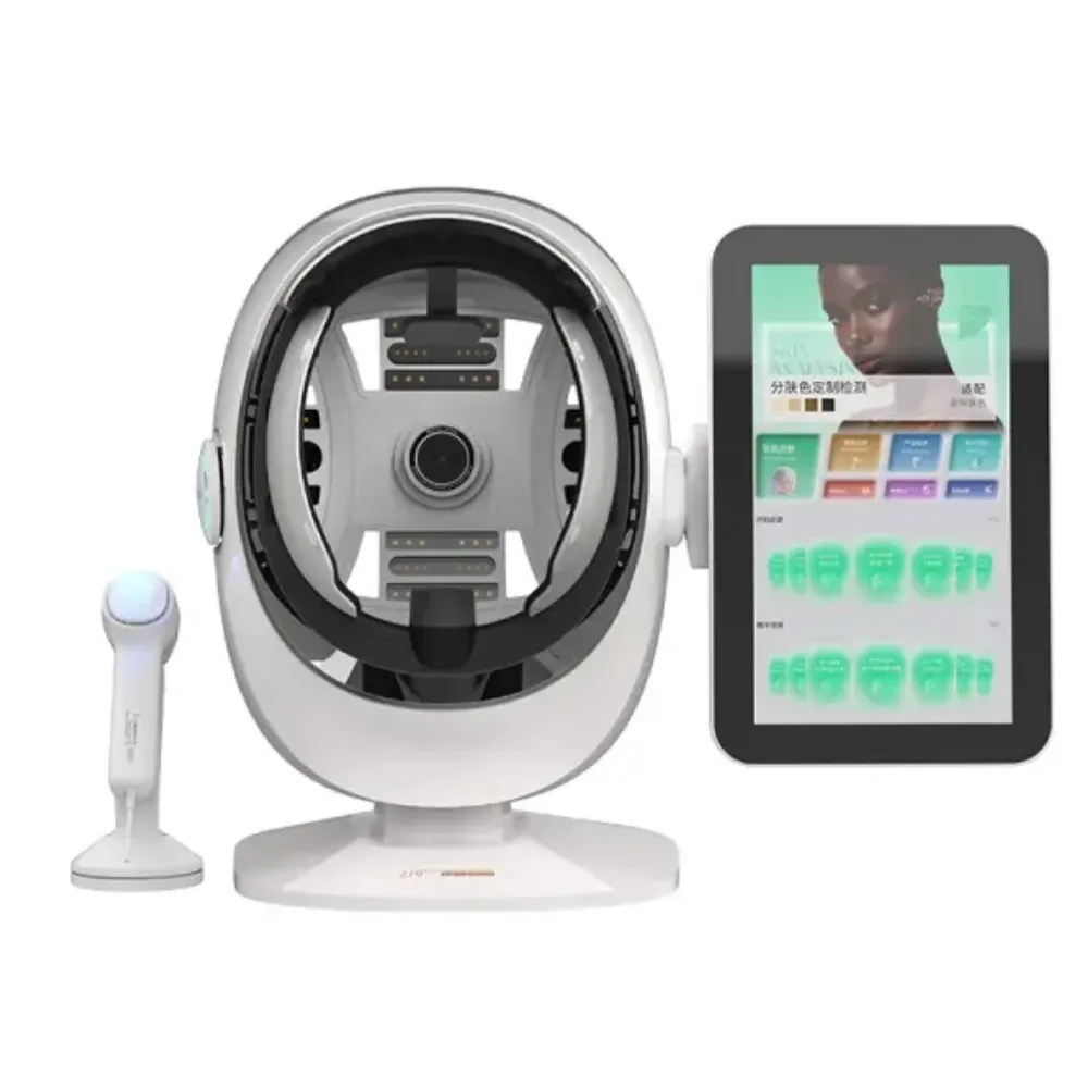
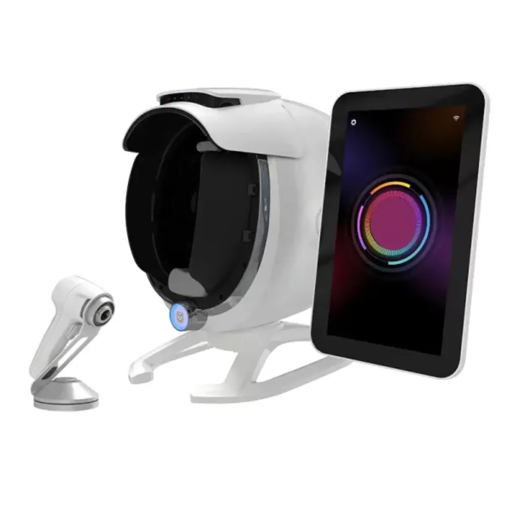
The K57 3D AI 8-spectral imaging system is one example of how these ideas come together in a single facial skin analyzer. Its design handles skin-tone differences through separate imaging channels and a controlled capture.
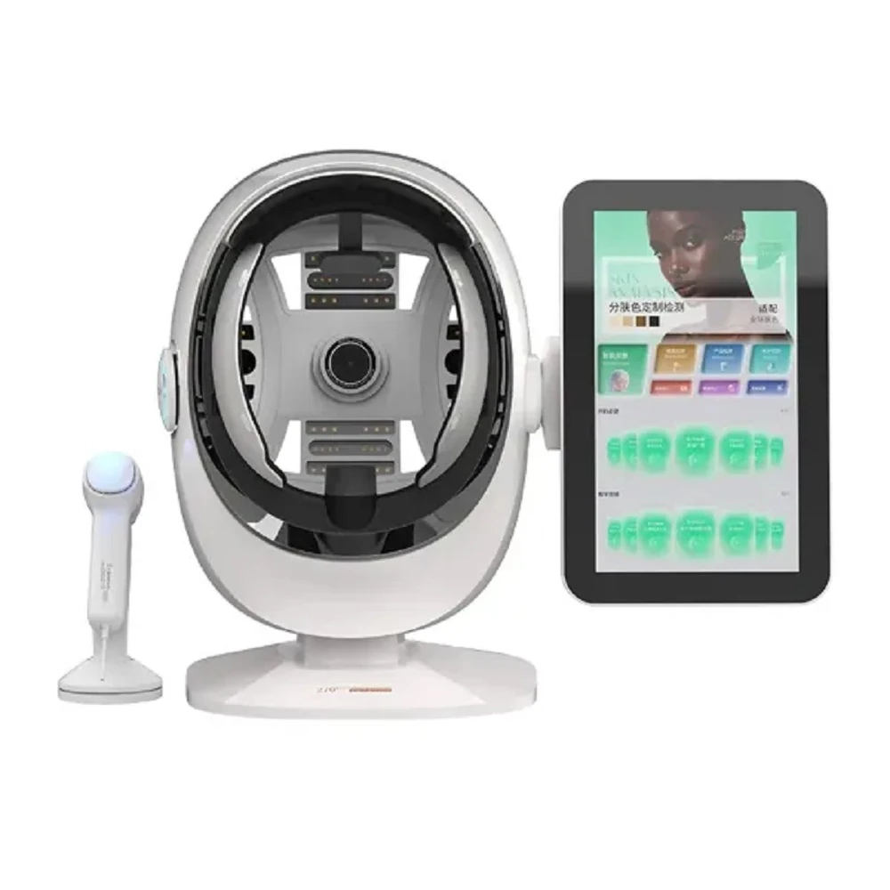
Separate pigment and blood channels are the basis for reading skin across different skin tones. Eight imaging channels are available. They include white-light RGB, parallel and cross polarization, Wood’s light (a long-established UV viewing method), UV, a hemoglobin channel, a melanin channel and a raw white-light reference image. Because the melanin and hemoglobin maps can be read side by side, a dark area can be identified as pigment or as vascular redness instead of being lumped together. Cross-polarized views suppress the glare from surface oil, which otherwise reads as dullness.
A semi-open shading hood, a forehead pad and a chin pad form a controlled dark space and lock the capture distance. The hood is not fully sealed, so strong side light, a direct spotlight or a bright window behind the client can still disturb the white-light and polarized channels. Clinics get more consistent results when the unit sits away from direct daylight and the room uses indirect lighting. Parameter calibration also lets a practice fine-tune the reading to its own baseline; when that option is switched off, the original data is shown as captured.
A 36 MP industrial-grade sensor records a single high-fidelity frame to hold fine surface texture and shallow pigment detail. One capture fires five flashes in sequence. It completes the front, left and right views across 270°, so the client is positioned once and never changes pose between light sources. Fewer repositioning steps mean fewer chances for the face to drift between the images that will later be compared.
Eighteen quantitative indicators cover moisture, pores, blackheads, sebum, barrier condition, acne, wrinkles, mixed and surface pigmentation, sun spots, porphyrins, collagen, roughness, deep and brown pigment, and sensitivity maps. Each one is graded A to E — green, blue, yellow, orange and red — and accompanied by a score, a grade, a count, an area and a percentage. A report that shows how much of the face is affected is far easier to discuss than a single headline number.
More than thirty functions sit alongside those indicators. The 16-type skin classification, a public four-axis model built on oily or dry, sensitive or tolerant, pigmented or non-pigmented, and wrinkled or firm, helps staff group clients consistently. A pH reference range of 4.5–6.5, skin-age estimation, aging simulation, sunscreen product testing and microscopic viewing for face and scalp are also included. Skin-age estimates and aging simulation are projections, so practitioners should read them as scenarios rather than measured values.
Images are uploaded for cloud analysis once the capture is complete. Reports can be opened through an H5 code, sent by email, downloaded as a PDF or printed directly. Client records stay in the archive, and horizontal, vertical and four-image comparisons let a clinic show the same area across two visits. Data center statistics summarize client numbers and concern distribution by month or year.
Because the reading depends on light, not on the face alone. Melanin absorbs across 400–700 nm, so deeper skin sends less light back to the camera. An exposure and a threshold that suit lighter skin will read a high melanin baseline as a pigmentation concern on deeper skin. Devices also differ in their channels, their light sources and their reference ranges, so the same feature can legitimately land on either side of a cut-off. Keeping the capture conditions consistent, then judging the result, keeps the comparison meaningful.
Ask for clean skin without makeup, and avoid scanning on the same day as exfoliation or an energy-based treatment. During the shot, the chin and forehead should rest on their pads, the nose should line up with the center guide, and the client should keep both eyes closed. Strong side light and direct sun from a window behind the chair are worth avoiding. For a follow-up, keep the same settings and roughly the same time of day; otherwise the longitudinal view will mix a real change with a change in conditions.
No. A skin analysis device produces images and reference values that help a practitioner explain what is visible and follow how it changes. It supports a skincare consultation and progress tracking; it is not a diagnostic device and it makes no medical claims. When a lesion looks unusual, or when a client reports symptoms rather than a cosmetic concern, the right next step is a physician. Keeping that boundary clear also makes the rest of the report more credible.
Most sessions follow the same short order: open or create the client record, capture the images, optionally take a moisture reading, then let the software build the report. How long that takes depends on how many views and add-on readings a practice chooses. Reports are commonly shared on screen, sent to the client by email, exported as a PDF or printed. What matters more than speed is that the same steps are repeated at every visit.
Yes, provided the conditions stay comparable. Client records and scan history are usually retained, and side-by-side views place two visits next to each other. The comparison is only useful when the same settings, the same channels and roughly the same lighting are used each time. If a score drops after the room was moved or the settings were changed, staff should treat the setup as the first suspect rather than celebrating a result.
Skin tone is not a flaw in the client and it is not a mystery in the machine. It is a variable that changes how much light returns, where the reference baseline sits, and whether pigment can be told apart from redness. Once a clinic understands those three mechanisms, a surprising score stops looking like an error and starts looking like a question about the setup.
The practical answer is unglamorous. Match the exposure before the shot, read pigment and blood through separate channels, hold the face still, and keep every follow-up under the same conditions. Devices that build those steps into one capture make the routine easier to keep, especially when a practice serves clients across several skin tones. A report is only as trustworthy as the conditions that produced it.
Request the full parameter list or arrange a live demonstration to see how the imaging channels perform on your own client mix.